
Fig. 5.
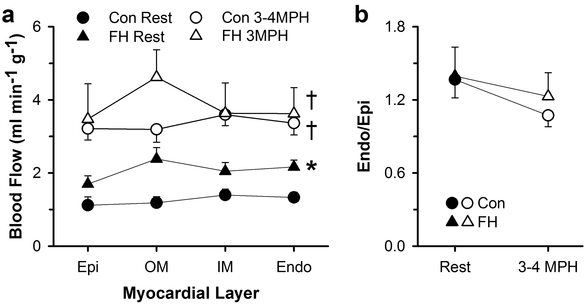
Myocardial blood flow per gram of myocardium in four layers across the left ventricular free wall at rest and during exercise in control and FH swine. (a) Blood flow to the subepicardium (Epi), outer mid (OM) myocardium, inner mid (IM) myocardium, and subendocardium (Endo) were determined by infusion of microspheres at rest and during treadmill exercise at 3–4 mph. (b) Subendocardial-to-subepicardial blood flow ratio (Endo/Epi) of the left ventricular free wall in control and FH swine at rest and during exercise at 3–4 mph. Values are mean ± SE, *p<0.05 versus Con Rest, †p<0.05 versus Rest within group.