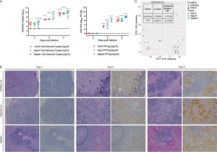
FIG 7.
EBOV-Makona infection results in viremia, histopathological changes, and large a transcriptional response in lymphoid tissue. (A) The mean ± SD of EBOV infectious virus titer and genome copy numbers were quantified in lymphoid tissues using a plaque assay and RT-qPCR for each animal. Significance was determined using one-way ANOVA with Dunnett’s multiple-comparison test relative to the limit of detection, which is represented as a dashed line. *, P < 0.05; ***, P < 0.001; ****, P < 0.0001. (B) Hematoxylin and eosin staining (left) and immunohistochemistry staining (right) (brown indicates reactivity) using anti-VP40-specific antibody of lymphoid tissues at 2, 4, and 6 DPI; magnification ×20. (C) Principal component analysis of lymphoid-specific DEGs over time.

