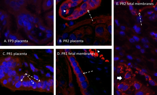
FIG 7.
(A) Panflavivirus IF (red, flavivirus; blue, DAPI) staining in the placenta of a ZIKV RNA-negative dam infected with the FP isolate demonstrating a lack of ZIKV IF in the villous placenta. (B and C) ZIKV IF was noted in occasional villous trophoblasts in placentas from dams infected with the PR isolate (white arrows), consistent with ZIKV infection of the syncytial layer. (D) Infection of the amnion epithelium was also observed (arrow). (E) ZIKV IF was also observed in the chorion/decidual layer of the fetal membranes in a dam infected with the PR isolate (large arrow). Autofluorescing red blood cells are indicated with arrowheads.

