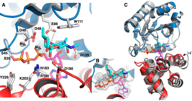
FIG 2.
Structure of ANT(9) in complex with spectinomycin (cyan), ATP (magenta), and magnesium (white). The N-terminal domain is in blue, and the C-terminal domain is in red. (A) Interactions of spectinomycin in the active site of ANT(9)-ATP-spc. Direct hydrogen bonds to spectinomycin are shown as dashed lines. (B) Fo-Fc omit maps of spectinomycin, ATP, and magnesium contoured at 2.5σ. (C) Superposition of the N-terminal domain of ANT(9) apo- (gray) with ANT9-ATP-spc. Upon ligand binding, the C-terminal domain rotates 14° toward the N-terminal domain.

