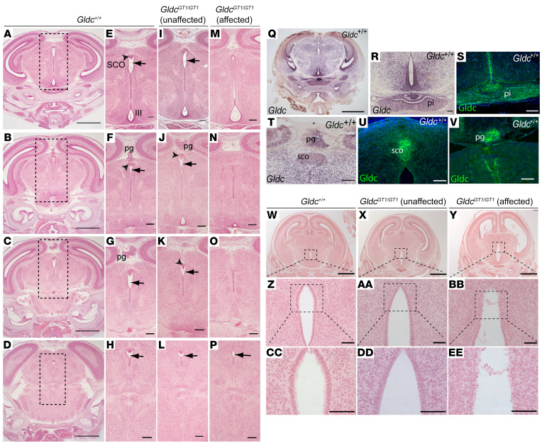
Figure 2. Ventriculomegaly is associated with aqueduct stenosis in Gldc-deficient fetuses.
Coronal sections in a rostral-caudal sequence (at levels shown in A–D in wild-type brain) show continuity of the aqueduct of Sylvius in Gldc+/+ (arrows in E–H) and unaffected GldcGT1/GT1 (I–L) fetuses at E18.5. In contrast, the aqueduct narrows and exhibits discontinuities in GldcGT1/GT1 mutants with ventriculomegaly (affected) (M–P). Boxed areas in A–D show enlarged regions in E–P. At E16.5, Gldc mRNA is widely expressed in the brain (Q, R, and T), with abundant expression in the pineal gland (pg), subcommissural organ (sco), and pituitary (pi). Immunohistochemistry confirms localization of Gldc protein at these sites at E18.5 (S, U, and V). The ependymal cell lining of the third ventricle (boxed in W–Y, enlarged in Z–EE) appears disrupted in GldcGT1/GT1 fetuses at E18.5 (Y). Scale bars: 0.1 mm (R–V and Z–EE), 0.5 mm (A–P), and 1 mm (Q and W–Y).

