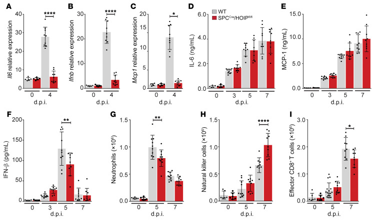
Figure 6. Loss of HOIP from the alveolar epithelium inhibits alveolar epithelial-driven inflammatory response to IAV infection.
(A–G) WT and SPCCre/HOIPfl/fl mice were infected with a lethal dose of WSN. (A–C) AT2 cells at 0 and 4 d.p.i. (n = 9) analyzed for Il6 mRNA (A), Ifnb mRNA (B), and Mcp1 mRNA (C). (D–F) BALF analyzed by ELISA at 0, 3, 5, and 7 d.p.i. (n = 9) for IL-6 (D), MCP-1 (E), and IFN-β (F). (G–I) Lung immune cell populations at 0, 5, and 7 (n = 9) d.p.i. analyzed by flow cytometry for Ly6G+CD11b+CD24+ neutrophils (G), NK1.1+CD11bhiCD24hi natural killer cells (H), and CD44+CD62L–CD8+ T cells (I). Means ± SD overlaid with individual data points representing replicates are depicted; *P < 0.05, **P < 0.01, ****P < 0.0001 (1-way ANOVA, Bonferroni post hoc test).

