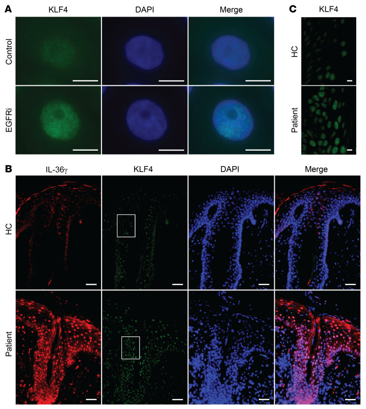
Figure 6. Increased KLF4 levels in vitro and in vivo upon EGFR inhibition.
(A) Representative images of KLF4 expression (green) in PHKs after erlotinib or control DMSO exposure for 24 hours. Nuclei were stained with DAPI. Scale bars: 10 μm. Data are representative of 3 independent experiments. (B and C) Immunofluorescent staining with mouse anti-KLF4 (green) and rabbit anti–IL-36γ (red) antibodies of formalin-fixed, paraffin-embedded skin sections of acneiform eruption patients and healthy controls (HC). Nuclei were stained with DAPI. The white-boxed regions in B were zoomed separately in C. Scale bars: 50 μm (B) and 10 μm (C). Pictures are representative of 5 patients and 3 healthy individuals.

