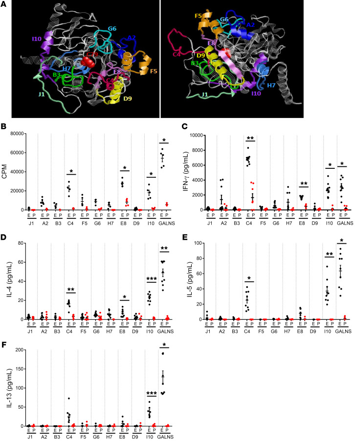
Figure 1. Selection of immunodominant peptides within the GALNS enzyme.
(A) Location of the synthetic peptides in the 3D structure of GALNS enzyme. Peptides A2 (blue), B3 (green), C4 (magenta), D9 (yellow), E8 (violet), F5 (orange), G6 (cyan), H7 (light blue), I10 (purple), and J1 (light green) are highlighted. The active site of the protein (C79) is shown in red. (B–F) Selection of immunodominant peptides after in vitro stimulation of splenocytes. MKC mice were treated with 16 intravenous weekly infusions of human GALNS (E, black dots) or PBS (P, red dots). Ten days after the last infusion, mice were euthanized and splenocytes were in vitro stimulated with GALNS or a single peptide. The background levels from unstimulated cells were subtracted. (B) Levels of splenocyte proliferation (n = 6 measurements from 2 different mice) and secretion levels of cytokines (C) IFN-γ, (D) IL-4, (E) IL-5, and (F) IL-13 (n = 9 measurements from 3 different mice). Data are shown as scatter plots with mean ± 95% CI. *P < 0.05; **P < 0.01; ***P < 0.001 represent statistically significant differences between treated and untreated mice as determined by 2-tailed paired t test.

