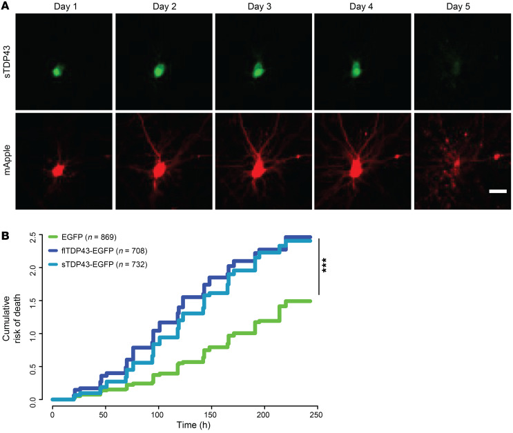
Figure 5. sTDP43 overexpression is neurotoxic.
(A) Example of a single neuron expressing mApple and sTDP43-EGFP, tracked by longitudinal fluorescence microscopy. Fragmentation of the cell body and loss of fluorescence indicates cell death. (B) The risk of death was significantly greater in neurons overexpressing sTDP43-EGFP and flTDP43-EGFP, compared with those expressing EGFP alone. EGFP n = 869, flTDP43-EGFP n = 708, sTDP43-EGFP n = 732, stratified among 3 replicates. ***P < 2 × 10–16 by Cox proportional hazards analysis. Scale bar: 20 μm (A).

