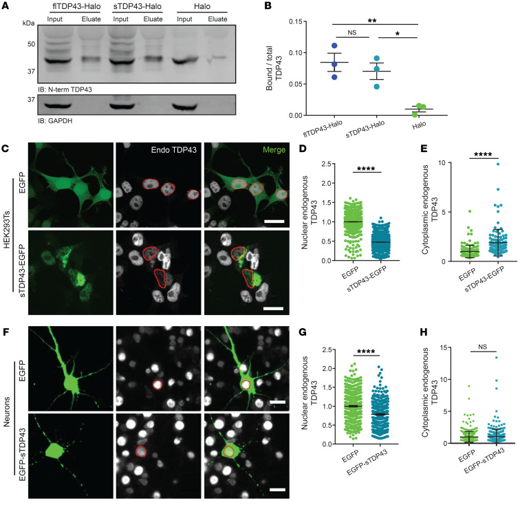
Figure 6. sTDP43 overexpression leads to the cytoplasmic deposition and nuclear clearance of endogenous TDP43.
(A) HaloTag fusions of flTDP43 or sTDP43 were expressed in HEK293T cells and immunoprecipitated with HaloLink. Bound TDP43 was immunoblotted (IB) with a C-terminal TDP43 antibody. GAPDH served as a loading control. (B) Quantification of data shown in A, demonstrating the fraction of total TDP43 bound to flTDP43-Halo, sTDP43-Halo, or Halo alone. Data were combined from 3 replicates. *P < 0.05, **P < 0.01 by 1-way ANOVA with Dunnett’s post hoc test. (C) HEK293T cells were transfected with EGFP or EGFP-tagged sTDP43, and then immunostained using an antibody that recognizes the endogenous TDP43 C-terminus (Endo). Red, nuclear regions of interest (ROIs) determined by DAPI staining. (D) Nuclear, endogenous TDP43 is reduced by sTDP43 overexpression in HEK293T cells. EGFP n = 1537, sTDP43-EGFP n = 1997, 3 replicates. ****P < 0.0001 by 2-tailed t test. (E) Cytoplasmic endogenous TDP43 is elevated by sTDP43 overexpression in HEK293T cells. EGFP n = 129, sTDP43-EGFP n = 113, 3 replicates. ****P < 0.0001 by 2-tailed t test. (F) Primary mixed rodent cortical neurons were transfected with EGFP or EGFP-tagged sTDP43, and then immunostained using a C-terminal TDP43 antibody. Red, nuclear ROIs determined by DAPI staining. (G) sTDP43 overexpression resulted in a significant drop in nuclear, endogenous TDP43 in primary neurons (EGFP n = 395, EGFP-sTDP43 n = 323, 3 replicates; ****P < 0.0001 by 2-tailed t test), but this was not accompanied by increases in cytoplasmic, endogenous TDP43 (H) (EGFP n = 394, EGFP-sTDP43 n = 323, 3 replicates, NS by 2-tailed t test). Scale bars: 20 μm (C and F).

