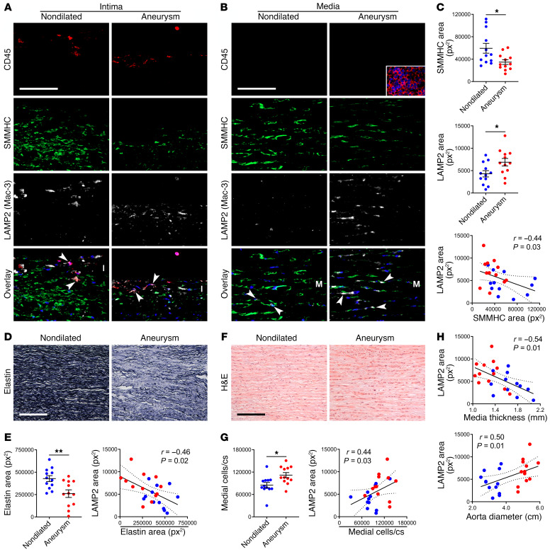
Figure 10. Degradative SMCs in human TAAD.
Ascending aortas were procured from 24 subjects undergoing aortic surgery (Aneurysm) or from organ donors (Nondilated). (A and B) Immunofluorescence analysis for CD45 (red), SMMHC (green), LAMP2 (also known as Mac-3; white), and overlays with DAPI-labeled nuclei (blue) showing LAMP2+ leukocytes (arrows) in the intima (I) and LAMP2+ SMCs (arrows) in the media (M); inset of spleen positive control (scale bars: 100 μm). (C) Expression of SMMHC and LAMP2 (n = 12) and correlation of SMMHC to LAMP2 (n = 24). px2, pixels squared. (D) Verhoeff stain of aortic media for elastin (scale bar: 200 μm). (E) Elastin expression (n = 12) and correlation of elastin loss to LAMP2 (n = 24). (F) H&E stain of media (scale bar: 200 μm). (G) Number of medial cells per cross section (cs) (n = 12) and correlation of medial cells to LAMP2 (n = 24). (H) Correlation of media thickness and aorta diameter to LAMP2 (n = 24). Data are represented as individual values with mean ± SEM bars or linear regression lines. *P < 0.05, **P < 0.01 by t test (C, E, and G) or Spearman’s test for r correlation coefficient (C, E, G, and H).

