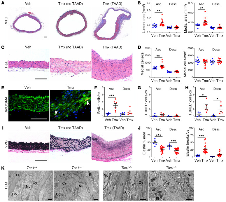
Figure 2. Aortic pathology is characterized by SMC proliferation and elastin fragmentation.
Tsc1fl/fl Myh11-CreERT2 mT/mG mice were treated with tamoxifen (Tmx) or vehicle (Veh) at 1.5 weeks of age and their ascending (Asc) and proximal descending (Desc) thoracic aortas were examined by histology at 12 weeks of age; representative photomicrographs of ascending aortas without or with TAAD are shown. Scale bars: 100 μm. (A) Masson’s trichrome (MTC) stains and (B) lumen and media area (n = 9–17). (C) H&E stains and (D) number of medial cells per cross section (cs) or area (n = 9–17). (E) BrdU reactivity (red color marked by arrows), SMA expression (green color), and DAPI-labeled nuclei (blue color) in subset of mice receiving BrdU for 2 weeks and (F) number of BrdU+ medial cells per cross section (n = 6). Number of TUNEL+ medial cells per cross section at (G) 12 weeks and (H) 24 weeks (n = 5). (I) Verhoeff–Van Gieson (VVG) stains and (J) elastin fraction of media area and number of elastin breaks per cross section at 12 weeks (n = 6–21). Data are represented as individual values with mean ± SEM bars. *P < 0.05, **P < 0.01, ***P < 0.001 for Tmx vs. Veh by 2‑way ANOVA. (K) Transmission electron microscopy (TEM) of ascending aortas from tamoxifen-treated Tsc1fl/fl Myh11-CreERT2 mT/mG (Tsc1−/−) and Myh11-CreERT2 mT/mG (Tsc1+/+) mice at 24 weeks showing elastin attenuation, loss of cytoplasmic filaments, and more cytoplasmic organelles extending from perinuclear region to periphery. El, elastin; Co, collagen; Nu, nucleus; Cy, cytoplasm. Scale bars: 1 μm.

