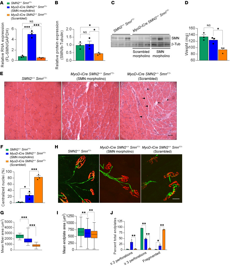
Figure 8. SMN repletion mitigates muscle defects in symptomatic mutants.
Levels of the (A) SMN2-derived FL-SMN transcripts and (B) SMN protein in gastrocnemius muscles of controls and mutants either administered an SMN2 splice-switching MO or a nonspecific MO at 7 months of age; 1-way ANOVA, n = 3 mice of each genotype. (C) Western blot probed for SMN protein in skeletal muscle after treatment with SMN-MO or scrambled MO. (D) Muscle weights in mice treated as described in the previous panels; 1-way ANOVA, n ≥ 3 mice in each group. (E) Transverse sections of gastrocnemius muscles from treated mutants and controls. Less pathology in the form of central nuclei (arrowheads), hypotrophic fibers (open arrows), and degenerating fibers (solid arrows) observed in SMN-MO treated muscle. Scale bar: 100 μm. Quantified results of (F) central nuclei and (G) myofiber areas in muscles from the mice; 1-way ANOVA, n ≥ 300 fibers from n ≥ 3 mice of each cohort. (H) Endplates from the 3 sets of mice; fewer fragmented NMJs are seen in the SMN-MO treated mutant than in its counterpart administered scrambled MO. Scale bar: 25 μm. (I) NMJ area measurements and (J) NMJ complexity analysis in gastrocnemius muscles of treated mutants or littermate controls; 1-way ANOVA, n ≥ 400 endplates from n ≥ 3 mice of each genotype for results in I and J. *P < 0.05; **P < 0.01; ***P < 0.001.

