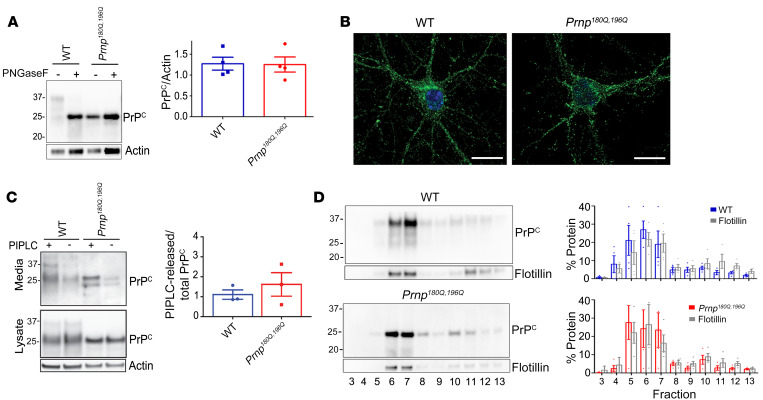
Figure 1. PrP180Q/196Q traffics similarly to WT PrPC in primary neurons and in mice.
(A) Representative Western blot of PNGase-F–treated brain extracts from age-matched Prnp180Q/196Q and WT mice reveal similar PrPC expression levels (quantified in right panel) (100–250 day old mice); n = 4/group. (B) PrP immunocytochemistry shows that unglycosylated PrP180Q/196Q traffics to neuronal processes in primary cortical neurons, as does PrP in WT neurons; n = 3 experiments. Scale bars: 10 μm. (C) Representative Western blots of phospholipase C–cleaved (PIPLC-cleaved) PrP180Q/196Q and WT PrP from the surface of cortical neurons show that surface PrPC levels are similar (media); n = 3 experiments. The additional band in the media (~23 kDa) may be a cleaved form of PrP. (D) PrP180Q/196Q and WT PrPC, together with flotillin, localize to detergent-resistant membranes in the brain; n = 3/group. Unpaired, 2-tailed Student’s t test, no significant differences (A and C).

