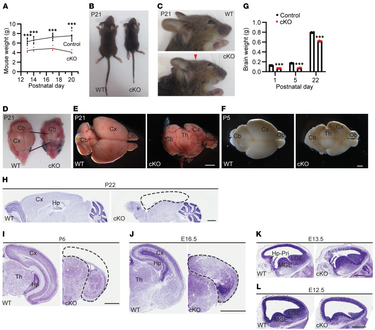
Figure 1. Conditional Kat8 deletion causes early lethality and cerebral hypoplasia.
(A) Growth curves for control and homozygous knockout (cKO) mice (n = 14 and 4, respectively). (B) Photos of wild-type and cKO mice at P21. (C) Enlarged photos of head parts of the mice shown in B. The “flat-head” phenotype refers to the flat head surface above the cerebrum (indicated with a red arrowhead). (D) Photos of deskinned heads from the mice shown in B. (E) Brain images for the wild-type and cKO mice shown in B. (F) Representative brain images for the wild-type and cKO mice at P5. See Supplemental Figure 2, C–F, for brain images of another pair at P5 and 3 pairs at P1, E18.5, and E16.5. (G) Brain weight at P1, P5, and P22 (n = 5, 3, and 4 for each genotype, respectively). (H and I) Nissl staining of sagittal (H) or coronal (I) brain sections at P22 or P6. (J–L) Nissl staining of coronal (J) or sagittal (K–L) embryo sections at E16.5, E13.5, or E12.5. For panels H–L, mainly the cerebrum or its precursor is shown. Dashed lines demarcate the cerebral cortex. The mutant cortex is largely lost at P6 (I) and P22 (H). Neocortical and hippocampal lamination is not evident in the mutant at E16.5 (J). The small mutant LGE at E12.5 is due to section orientation. For (B–F), images are representative of 5 pairs of wild-type and mutant mice, and for (H–L), each image is representative of 3 experiments. For A and G, unpaired 2-tailed Student’s t tests were used; ***P < 0.001. Scale bars: 2 mm (E), 1 mm (F and H–J), and 0.5 mm (K–L). Cb, cerebellum; CP, cortical plate; Cx, cerebral cortex; Hp, hippocampus; Hp-Pri, hippocampal primordium; LGE, lateral ganglionic eminence; MGE, medial ganglionic eminence; Ob, olfactory bulb; Th, thalamus.

