Figure 1.

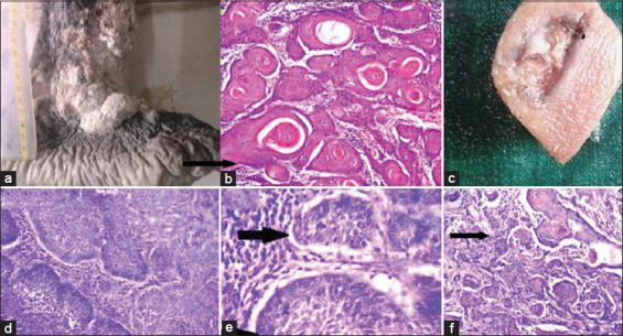
(a) Gross photograph of squamous cell carcinoma (SCC) showing large ulceroproliferative growth over the right leg. (b) Well-differentiated SCC – photomicrograph showing tumor cells with keratin pearls (black arrow) (hematoxylin and eosin [H and E], × 100). (c) Gross appearance of basal cell carcinoma showing ulcerative growth. (d and e) photomicrograph showing clefting artifact (black arrow) and peripheral palisading of tumor cells (arrow head) (H and E, × 400). (f) Basosquamous carcinoma – photomicrograph showing areas of squamous differentiation (black arrow) (H and E, × 100)
