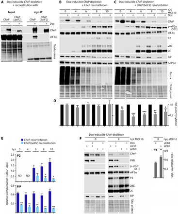
Fig. 3. The effects of CReP depletion on BiP/PVSRIPO translation are eIF2α dependent.

(A) Cells with combined dox-inducible CReP depletion and WT CReP/CReP(ΔeIF2) reconstitution were treated with dox (36 hours) before lysis and IP with anti-Myc beads. Lysates were compared by immunoblot to assess eIF2α binding. (B to E) HeLa cells with endogenous CReP depletion coupled with WT CReP (B) or CReP(ΔeIF2) (C) reconstitution were dox-induced, infected with PVSRIPO, and analyzed by immunoblot and puromycylation assay as described in Fig. 1. ND, not detected. (F) Cells with dox-inducible CReP depletion were mock- or dox-treated (36 hours), transfected with control siRNA or siRNA targeting PKR (36 hours), infected with PVSRIPO, and lysed for immunoblot analysis at the indicated intervals. (D to F) Statistical significance was assessed by Student’s two-tailed t test comparison at each time point between −/+ dox at each time point (D), relative compensation between the two cell lines [WT CReP versus CReP(ΔeIF2)] (E), or −/+ siRNA targeting PKR (F) for the indicated data (bar graphs represent mean and SEM; n = 3); *, **, *** corresponds to P < 0.05, 0.005, and 0.0005, respectively).
