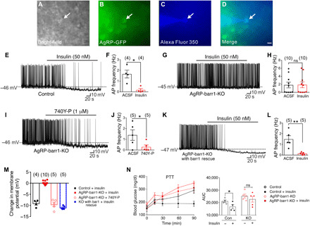
Fig. 4. Barr1 deficiency impairs insulin action on AgRP neurons.

(A to M) Electrophysiological studies with AgRP neurons from 8- to 12-week-old male mice maintained on standard chow. (A to D) Bright-field (A) or fluorescein isothiocyanate (GFP) illumination (B) of an AgRP neuron from AgRP-Z/EG reporter mice. (C) Complete dialysis of Alexa Fluor 350 from the intracellular pipette. (D) Merged image of a targeted AgRP neuron (arrow). Scale bar, 50 μm. (E to H) Current-clamp recording depicting insulin-induced hyperpolarization and reduced action potential (AP) firing frequency of control AgRP neurons (E and F) and lack of these responses in barr1-deficient AgRP neurons (G and H). ACSF, artificial cerebrospinal fluid. (I and J) The PI3 kinase activator 740Y-P elicits insulin-like responses in barr1-deficient AgRP neurons. (K and L) Reexpression of barr1 in AgRP neurons of adult AgRP-barr1-KO mice restores normal insulin responses. (M) Summary of the effects of insulin and 740Y-P on the membrane potential of AgRP neurons. (N) Central administration of insulin (200 μU into the lateral ventricle) fails to inhibit pyruvate (2 g/kg i.p.)–induced hyperglycemia in AgRP-barr1-KO mice (HFD for 12 weeks; 20-week-old males; n = 4 or 5). Data are given as means ± SEM. *P ≤ 0.05; **P ≤ 0.01 [two-tailed Student’s t test (E to L) and one-tailed Student’s t test (N)]. ns, no significant difference.
