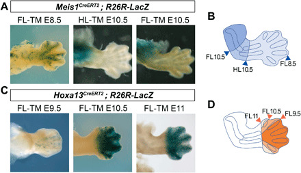
Fig. 3. Meis expression does not adjust to limb PD segmental borders.

(A) Lineage tracing of Meis1CreERT2-labeled cells by tamoxifen (TM) injection at different stages (TM.E8.5, n = 11; TM.E10.5, n = 3). (B) Lineage tracing of Hoxa13CreERT2-labeled cells by tamoxifen injection at different stages (TM.E9.5, n = 7; TM.E10.5, n = 4; TM.E11, n = 4). (C and D) Schemes showing the boundaries of the regions colonized by Meis-expressing cells (C) and Hoxa13-expressing cells (D) at different labeling time points. Besides minor leakiness observed following injections at E9.5 (C), the lineage of Hoxa13-expressing cells respects the zeugopod-autopod boundary.
