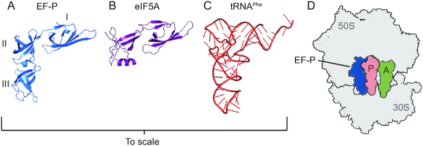
Figure 3.
The three dimensional structures of EF-P, eIF5A, and tRNA are similar. A) Three dimensional structure of Thermus thermophilus EF-P (PDB ID 1UEB). Domains I, II and III are labeled accordingly. B) Three dimensional structure of S. cerevisiae eIF5A (PDB ID 3ER0). C) Three dimensional structure of S. cerevisiae tRNAPhe (PDB ID 1EVV). D) The location of EF-P (blue) bound to a ribosome (grey) stalled at a polyproline sequence solved by cryo-electron microscopy (PDB ID 6ENJ). The positions of the 50S subunit, 30S subunit, P-site tRNA (salmon) and A-site tRNA (green) are labeled accordingly.

