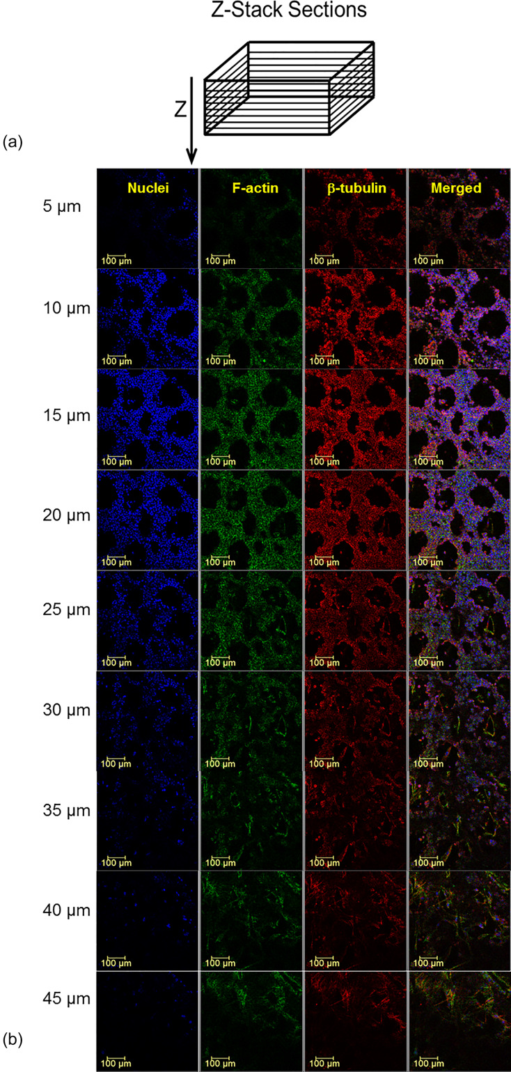
FIG. 3.
Successive confocal microscopy images of the endometrial barrier model. (a) Scheme of the cross sections perpendicular to the Z-axis. (b) The cross sections at indicated depths starting from the epical end of the endometrial layer. Antibodies: for F-actin, Cytopainter Phalloidin-iFluor 488 Reagent (ab176753), 1:1000 in DPBS 1% BSA; for β-tubulin, recombinant anti-beta tubulin antibody conjugated [EPR16774] (Alexa Fluor® 555) (ab206627) diluted with DPBS 1:500; and for nuclei, DAPI (D9542).

