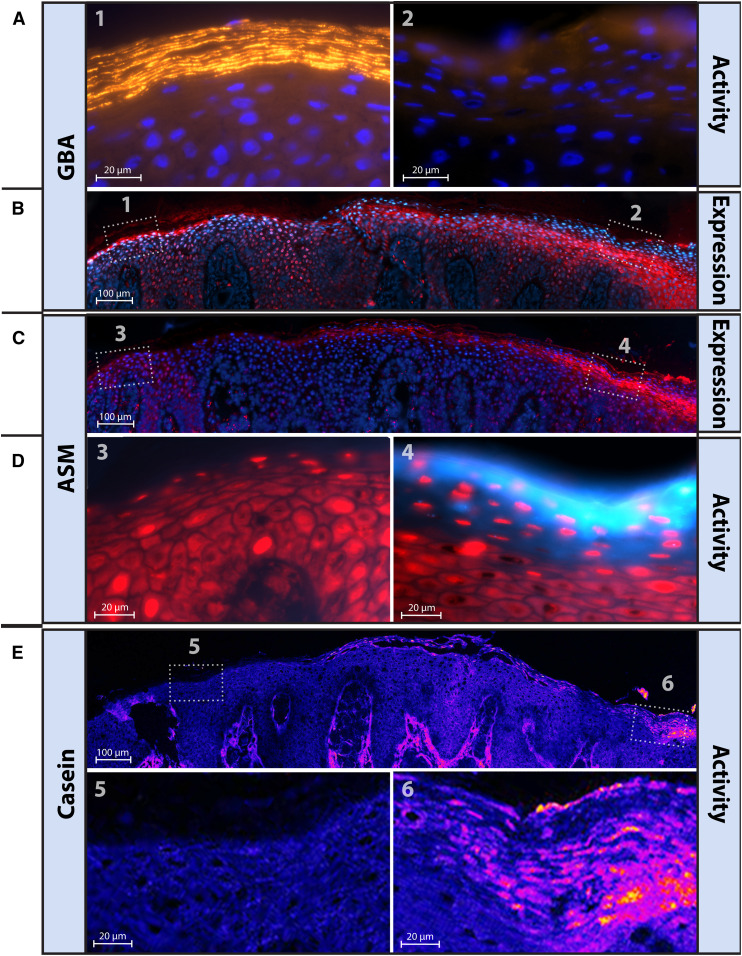
Fig. 5.
High local variation in GBA, ASM, and protease staining and/or activity in NTS patients. Five different stainings on five sequential 5 μm cut cryo-frozen sections from NTS7 and presented with matching skin areas. A: GBA-activity (yellowish, 63× magnification) from two different areas (areas 1 and 2) within a single cut section (blue, DAPI counterstaining). B, C: Respectively, GBA and ASM expression, labeled in red (with DAPI as blue counterstaining, magnification 20×). D: ASM activity (light blue, 63× magnification) from two different areas (areas 3 and 4) within a single cut section (red, propidium iodide solution (PI) counterstaining). E: Serine protease activity is shown in purple/orange (63× magnification) including a magnified area illustrating active serine proteases at some areas in the SC lipid layers.

