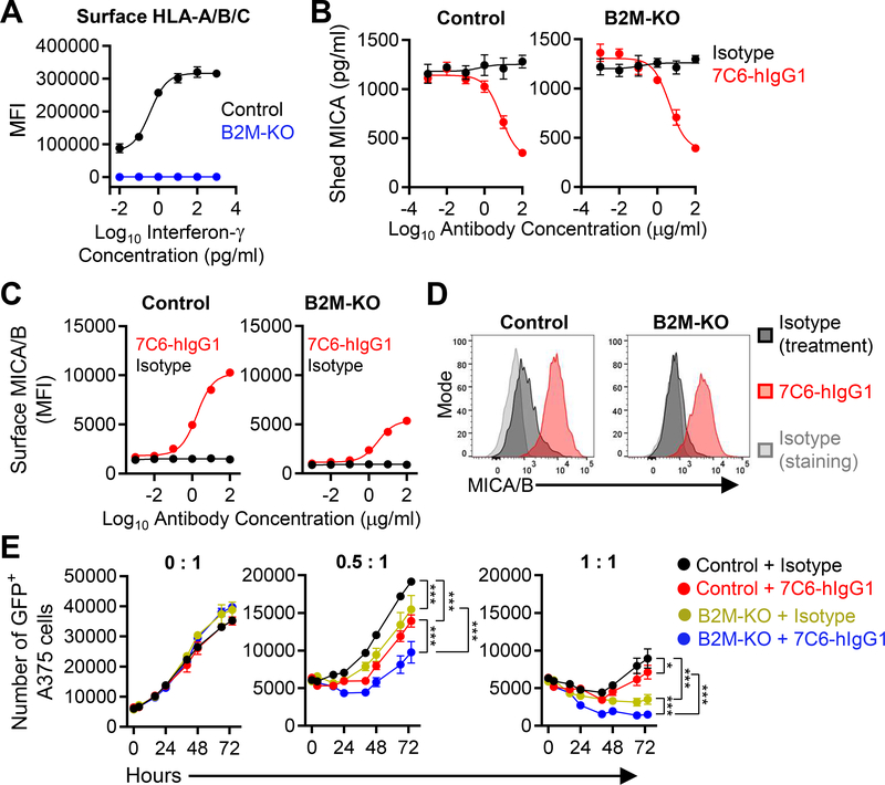
Figure 1. Inactivation of the B2M gene enhances NK cell–mediated killing of human melanoma cells in the presence of a MICA/B mAb.
(A) Validation of efficiency of B2M gene inactivation. Control or B2M-KO human A375 melanoma cells were treated with IFNγ for 24 hours and surface expression of HLA-A/B/C was quantified by flow cytometry. MFI = Mean Fluorescence Intensity. (B-D) Control or B2M-KO A375 cells were cultured for 24 hours with MICA/B (7C6-hIgG1) or isotype control antibodies at the indicated concentrations. (B) Quantification of shed MICA released by melanoma cells by sandwich ELISA. (C) MICA/B surface protein on control and B2M-KO melanoma cells was quantified by flow cytometry using PE-conjugated MICA/B mAb 6D4 or an isotype control mAb. (D) Histograms representative of the experiment shown in (C). (E) Effect of human NK cells on A375 cells dependent on MHC-I expression and MICA/B mAb treatment. GFP+ A375 cells (control or B2M-KO) were plated at a density of 5×10³ cells per well in a 96-well plate and pre-treated with 7C6-hIgG1 or isotype control mAbs (20 μg/mL) for 24 hours prior to addition of purified human NK cells at different effector to target ratios (0:1, 0.5:1 or 1:1). IL-2 (300 U/mL) was added to support NK-cell survival. The number of GFP+ A375 cells was quantified by imaging cytometry using a Nexcelom Celigo instrument at multiple timepoints over a 72-hour period. Data representative of three independent experiments (A-E). Statistical analyses were performed by two-way ANOVA with Bonferroni’s multiple comparison test (E). Error bars represent standard error (SEM) (A-C and E). *p<0.05, ***p<0.001.

