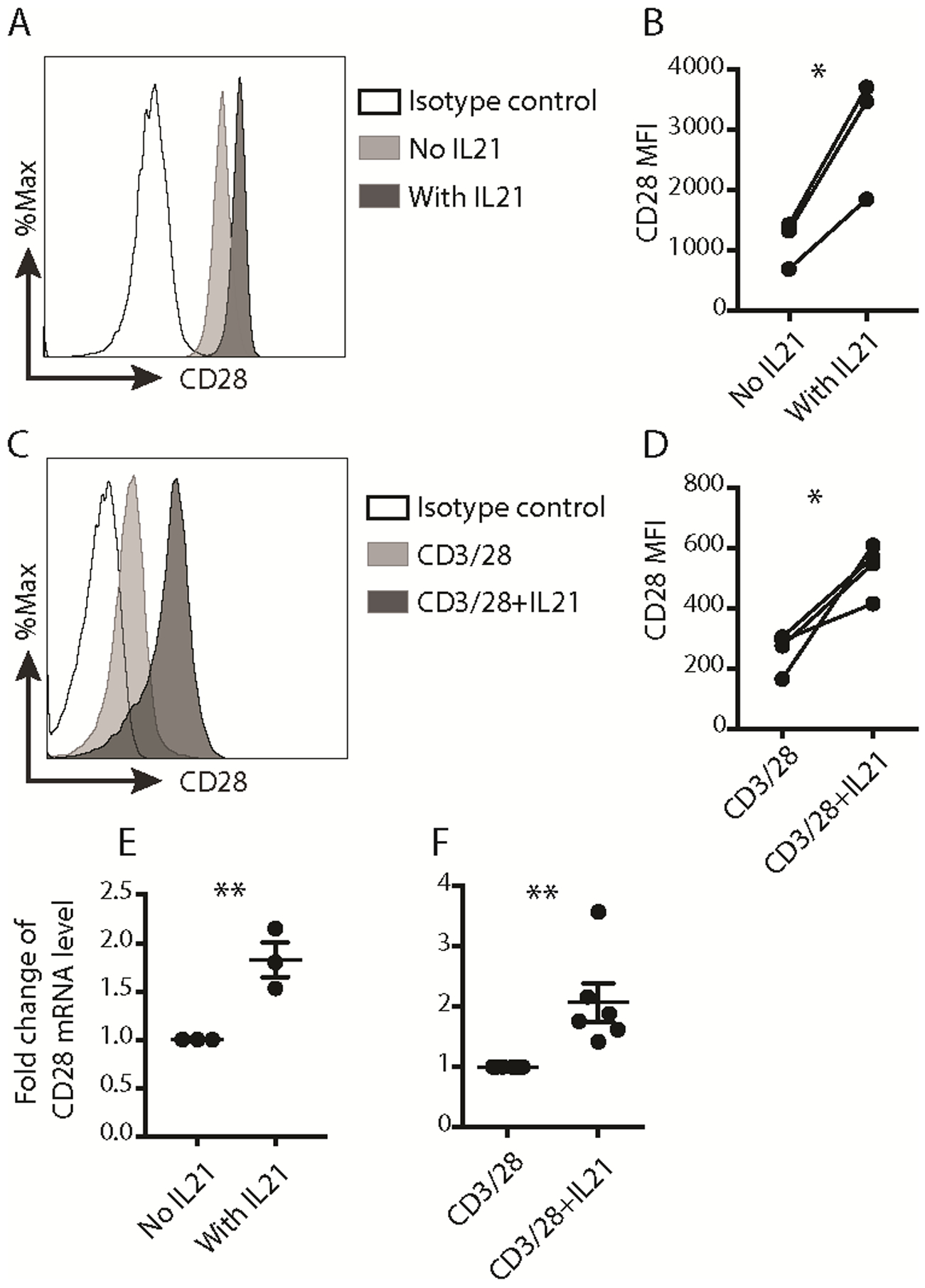
Fig. 1. IL21 upregulated CD28 expression in activated human naïve CD8+ T cells.

(A) Representative histogram of CD28 surface level on M27-specific CD8+ T cells and the isotype control antibody was used as a negative staining control. (B) MFI of CD28 protein expression on the surface of M27-specific CD8+ T cells. (n=3, * p < 0.05, paired t test). MFI: mean fluorescence intensity. (C) Representative histogram of CD28 surface expression on human naïve CD8+ T cells stained on day 7 after activation. The isotype control antibody was used as a negative staining control. (D) MFI of CD28 protein expression on the surface of CD8+ T cells activated with the indicated conditions for 7 days. (n=4, * p < 0.05, paired t test). (E) The quantitative RT-PCR results of CD28 mRNA in sort-purified M27-specific CD8+ T cells generated with or without IL21. The expression in cells expanded without IL21 was set as 1. (n=3, mean ± SEM, ** p < 0.01, unpaired t test). (F) The quantitative RT-PCR results of CD28 mRNA in human CD8+ T cells activated with the indicated conditions for 7 days. The expression in cells activated with anti-CD3/CD28 beads for 7 days was set as 1. (n=6, mean ± SEM, ** p < 0.01, unpaired t test). Results of quantitative RT-PCR for CD28 gene were normalized to RPL13A. The results in A and C were representative out of 3 (A) or 4 (C) independent experiments using cells from different healthy donors. The results in B, D, E and F were pooled from 3 (B, E), 4 (D) or 6 (F) independent experiments using cells from different healthy donors.
