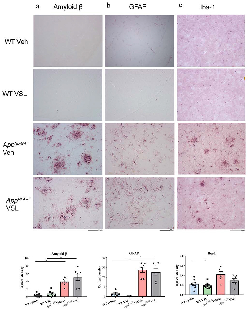
Fig. 7.
Effect of VSL#3 supplementation on Aβ accumulation and gliosis in brains of C57BL/6 (WT) and AppNL-G-F mice. Representative immunohistochemical staining for (a) Aβ (b) GFAP, and (c) Iba-1 are shown from temporal cortex. The representative images shown were taken at 20x. Quantitation of immunostaining was performed, and optical density values were averaged and shown in bar graphs. Data are represented as mean ± S.E.M. Significant differences were determined by two-way ANOVA, * p<0.05 (n=7), Scale bar 50μm.

