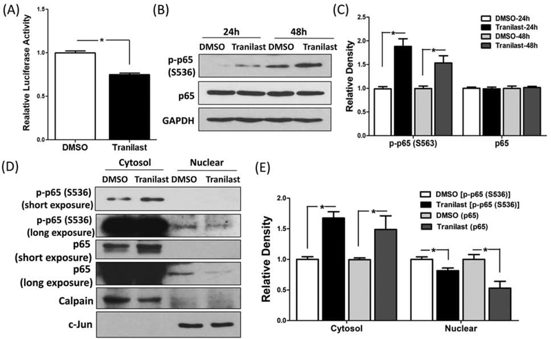
Figure 2.
(A) The level of RelA/p65 activity in LSMC transfected with a luciferase reporter construct containing preserved RelA/p65 binding sites and pRL-TK plasmid as control for transfection efficiency. Cells were treated with tranilast (200 μM) for 8 hrs. The ratio of Firefly:Renilla was determined and reported as relative luciferase activity as compared to control (DMSO) which was independently set as 1 (N=3). (B–C) Immunoblotting analysis of phosphorylated RelA/p65 at serine 536 and total RelA/p65 following treatment of LSMC with 200 μM tranilast for 24 hrs and 48 hrs (N=3). The relative band densities are shown in (C). (D) Shows protein levels of phosphorylated RelA/p65 at serine 536 and RelA/p65 in LSMC following treatment with 200 μM tranilast for 36 hrs (N=3). Cells were harvested and sub-fractionated into cytosol and nuclear proteins and subjected to immunoblotting analysis with Calpain and c-Jun serving as markers for the respective subcellular fractions. (E) The band density of phosphorylated RelA/p65 at serine 536 and RelA/p65 was semi-quantified and normalized to Calpain (cytosol) or c-Jun (nuclear). The results are presented as mean ± SEM of independent experiments as mentioned above using cells isolated from different patients in each set. P values (*P < 0.05) indicated by the corresponding lines.

