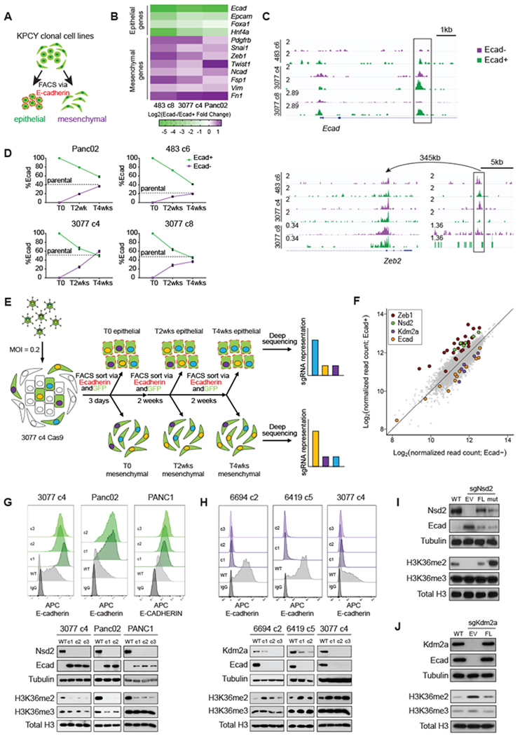
Figure 1: An epigenetic modifier-focused CRISPR-Cas9 screen identifies Nsd2 and Kdm2a as reciprocal regulators of epithelial-mesenchymal differentiation.

(A) Flow cytometry scheme for quantifying or sorting epithelial and mesenchymal subpopulations from clonal mouse pancreatic ductal adenocarcinoma (PDAC) cell lines. Epithelial subpopulations are identified by positive surface E-cadherin staining (Ecad+, outlined in red), while mesenchymal subpopulations are identified by the absence of surface E-cad (Ecad−).
(B) Heatmap summarizing triplicate qPCR experiments on sorted Ecad− and Ecad+ subpopulations for each of 3 cell lines. Increase and decrease in Ecad−/Ecad+ log2fold change is shown in purple and green, respectively.
(C) Representative ATAC-seq tracks of sorted Ecad− and Ecad+ subpopulations from 3 cell lines, shown in purple and green, respectively. Statistically significant (padj<0.1) enrichment peaks are boxed. Arrow indicates relationship between the Zeb2 promoter and a putative distal regulatory element.
(D) Summary of plasticity experiments performed on 4 cell lines. Sorted Ecad− and Ecad+ subpopulations were replated and passaged over the course of 4 weeks. Samples were reassessed for Ecad+% by flow cytometry every 2 weeks. Data are means ± SEM from biological replicates (n=3). Dotted lines represent the Ecad+% of the bulk, unsorted parental population of each cell line.
(E) Experimental outline for focused CRISPR screen. See materials and methods for a full description.
(F) sgRNA representation in Ecad− (x-axis) and Ecad+ (y-axis) populations as log2-transformed normalized read counts.
(G-H) Top: Representative Ecad flow histograms for clones expressing sgNsd2 (G)and sgKdm2a (H) compared to Cas9-only wildtype (WT) controls, as well as IgG-staining control for 3 cell lines. Bottom: Corresponding Western blots of cell lysates (top panels) and acid-extracted histones (bottom panels) with antibodies against the indicated proteins.
(I-J) Western blots of cell lysates (top panels) and acid-extracted histones (bottom panels) of Cas9-only wildtype (WT) controls and sgNsd2 (I) and sgKdm2a clones (J) expressing empty vector (EV), full-length (FL), or E1099K NSD2 (mut).
