Fig. 3.

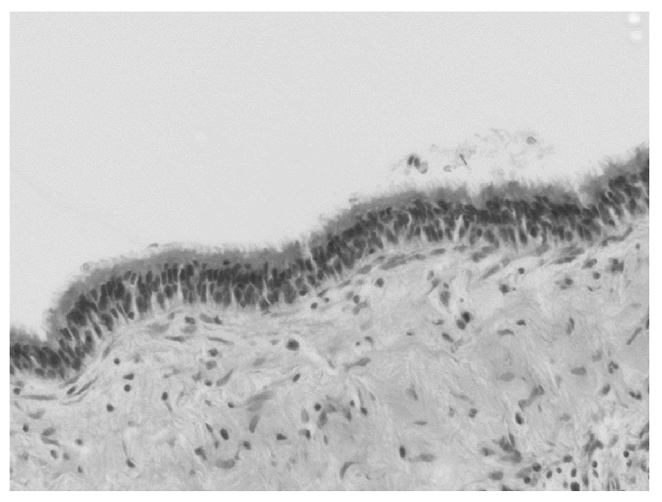
Histopathological examination showing a layer of ciliated columnar epithelium lining the inner side of the cyst, as well as acute inflammatory cells (hematoxylin & eosin stain, ×40).

Histopathological examination showing a layer of ciliated columnar epithelium lining the inner side of the cyst, as well as acute inflammatory cells (hematoxylin & eosin stain, ×40).