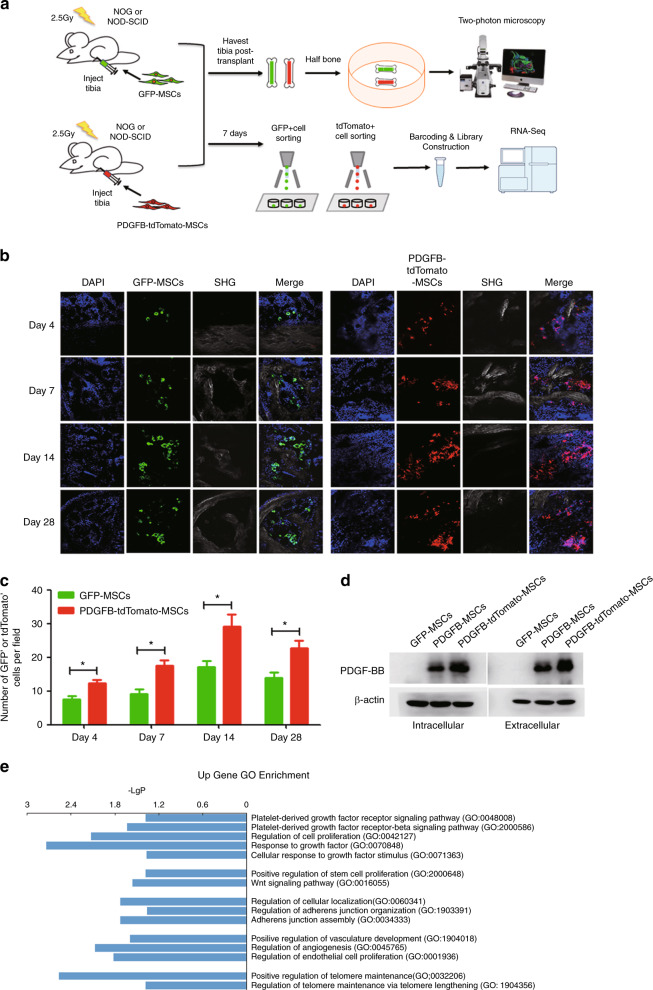
Fig. 5. PDGFB-MSCs proliferate more than GFP-MSCs in bone marrow cavity.
a Schematic representation of the experimental design. b Tracing of GFP+ and tdTomato+ cells by two-photon fluorescence microscopy at different time points after transplantation in the bone marrow cavity. c Quantification of the number of GFP+ and tdTomato+ cells per field in the injected tibias at multiple time points after transplantation. d Western blot of intracellular and extracellular PDGFB protein levels in GFP-MSCs, PDGFB-MSCs, and PDGFB-tdTomato-MSCs. e GO enrichment analysis of upregulated genes in PDGFB-tdTomato-MSCs harvested 1 week after transplantation (p < 0.05). SHG, Second harmonic generation. *p < 0.05.

