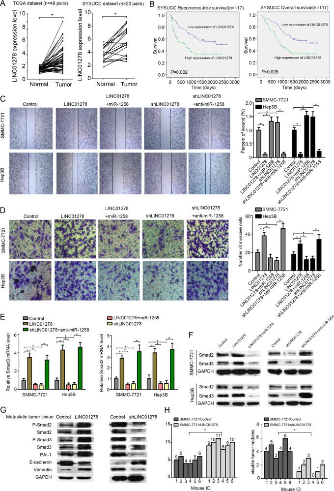
Fig. 4. LINC01278 increases HCC metastasis in vivo and in vitro.
a The expression of LINC01278 in paired HCC tissues and adjacent normal tissues in TCGA datasets (n = 49) and SYSUCC dataset (n = 20). b Correlation of recurrence-free survival (RFS) and overall survival (OS) and LINC01278 expression by Kaplan–Meier analysis in SYSUCC dataset. c The wound-healing assay in HCC cells. d The invasion assay in HCC cells. e The Smad2/3 mRNA expression of HCC cells transfected by LINC01278, shLINC01278, LINC01278+miR-1258, and shLINC01278+anti-miRNA-1258. f The protein expression of Smad2 and Smad3 in HCC cells transfected by LINC01278, shLINC01278, LINC01278+miR-1258, and shLINC01278+anti-miRNA-1258. g The protein levels of P-Smad2, Smad2, P-Smad3, Smad3, PAI-1, E-cadherin, and vimentin in metastasis tumor tissues. LINC01278, ectopic LINC01278 expression in HCC cells. h The lung metastasis of HCC cells transfected by LINC01278 and shLINC01278. shLINC01278, HCC cells were transfected by shRNA target LINC01278. miR-1258, HCC cells were transfected by miR-1258 expression vector. Anti-miR-1258, HCC cells were transfected by miR-1258 antisense plasmid. *P < 0.05.

