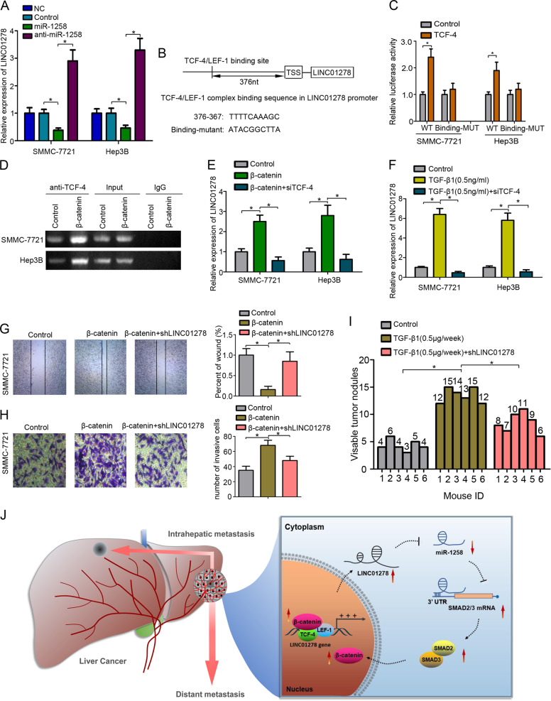
Fig. 5. LINC01278 was regulated by TCF-4.
a The expression of LINC01278 in HCC cells transfected by miR-1258 and anti-miR-1258. b The TCF-4/LEF-1 binding site in the LINC01278’s promoter sequence was identified. The mutant sequence was designed for the luciferase reporter assay. c The relative luciferase activity was detected in 293 T cells co-transfected by binding-WT/binding-mut and TCF-4. d ChIP assay was used to detect the TCF-4 to the TRE (TCF responsive element: TTCAAAG) regions in LINC01278 promoter. e The relative expression of LINC01278 in HCC cells transfected by β-catenin and siTCF-4. f The relative expression of LINC01278 in HCC cells treated with TGF-β1 (0.5 ng/ml) and siTCF-4. g, h The wound-healing assay and invasion assay in SMMC-7721 cells. i The lung metastasis of SMMC-7721 cells treated with TGF-β1 (0.1 µg/each time, three times a week) and shLINC01278. j Schematic illustration of “β-catenin/TCF-4-LINC01278-miR-1258-Smad2/3” axis. miR-1258, HCC cells were transfected by miR-1258 expression vector. Anti-miR-1258, HCC cells were transfected by miR-1258 antisense plasmid. TCF-4, HCC cells were transfected by TCF-4 expression vector. β-catenin, HCC cells were transfected by β-catenin expression vector. siTCF-4, HCC cells were transfected by siRNA target TCF-4. shLINC01278, HCC cells were transfected by shRNA target LINC01278. *P < 0.05.

