
Figure 1.
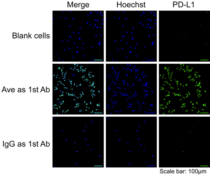
Confocal imaging of MDA-MB-231 breast cancer cell line after immunofluorescent staining. Sample groups: Hoechst, the nucleus stained by Hoechst; PD-L1, the PD-L1 expression; Ave as 1st Ab, the samples stained by avelumab as the primary antibody; IgG as 1st Ab, the samples stained by non-specific IgG as the primary antibody.