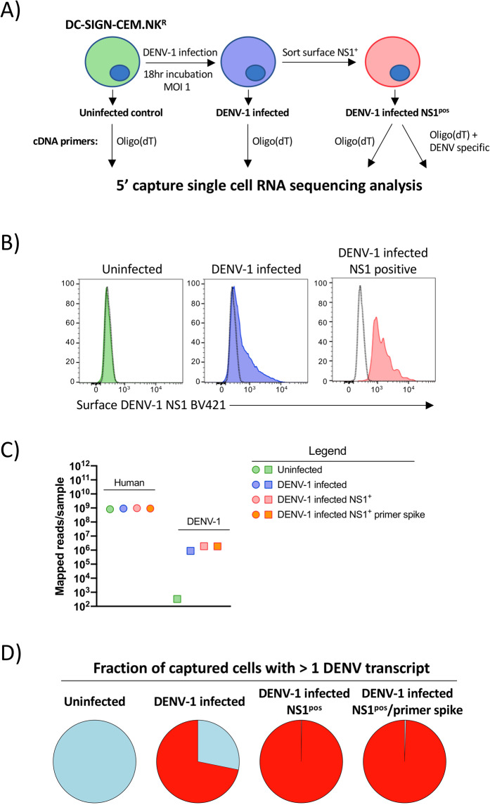
Figure 1.
5′ capture scRNAseq analysis of in vitro DENV infected cells. (A) Schematic representation of the infection, isolation, and analysis strategy utilized in this study. (B) Surface DENV-1 NS1 expression on uninfected, DENV-1 infected, and sorted NS1pos DENV-1 infected DC-SIGN expressing CEM.NKR cells utilized for scRNAseq analysis. (C) Number of PE reads confidently mapped from each sample to either the Gh38 human genome assembly or the DENV-1 (strain WestPac74) genome from each sample (D) Fraction of cells from each sample with > =1 confidently mapped DENV-1 transcript present.

