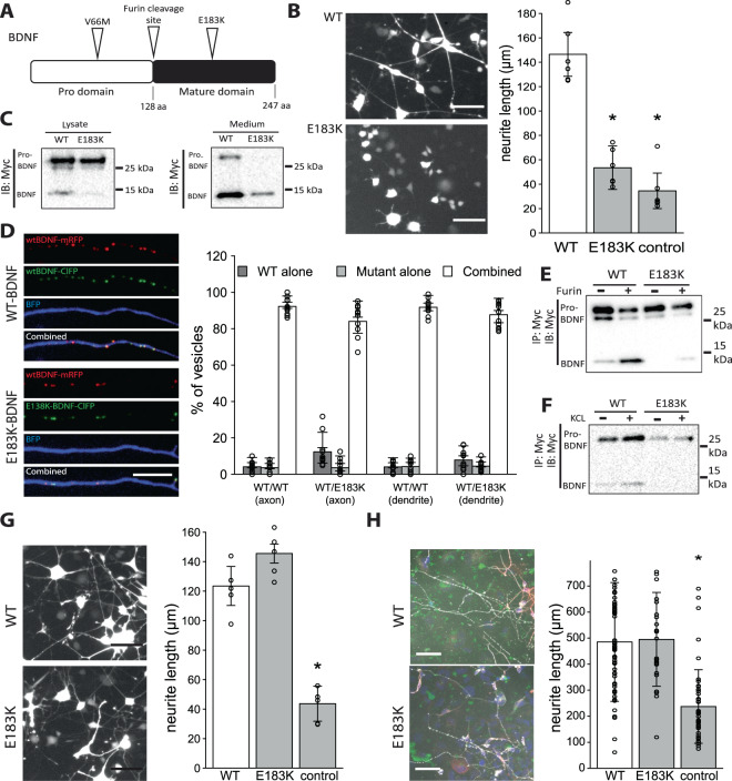
Figure 1.
Functional characterisation of a rare coding variant in BDNF (E183K). (A). Schematic representation of BDNF protein with the common variant (V66M) and rare variant (E183K) indicated. (B). PC12 cells were transfected with WT (top)/E183K (bottom) BDNF; neurite length was measured by fluorescence microscopy. Left panel: representative images from 3 experiments. Scale bar: 50 μm. Average neurite length per nucleus is shown (right panel; data point = mean of replicate); *p < 0.05, student’s t-test. (C). WT/mutant BDNF was transfected into PC12 cells and protein quantified by Western blot in cell lysate (left) and growth medium (right) using an antibody against a c-terminally fused myc-tag. (D). Cultured primary rat hippocampal neurons were co-transfected with RFP-tagged (red) WT BDNF and ClFP-tagged (green) WT BDNF (top image panel), or RFP-tagged WT and ClFP-tagged mutant (E183K) BDNF (bottom image panel). Co-localisation of the proteins was measured by fluorescent confocal microscopy in axons (shown here) and dendrites, and is presented as proportion of vesicles containing both (combined) or only one of the tagged proteins (center panel; data point = one axon). (Right panel: Density of dendritic BDNF positive vesicles containing either WT/WT BDNF or WT/E186K BDNF) Scale bar: 10 μm. (E). WT/mutant BDNF expressed in HEK293 cells was immunoprecipitated, followed by Furin-mediated protein cleavage. The cleavage products were analysed by Western blot. (F). WT/mutant BDNF were transfected into PC12 cells and depolarisation-dependent BDNF secretion triggered by addition of KCl. Amounts of secreted BDNF were measured by Western blotting. (G). TrkB-expressing PC12 cells were transfected with GFP and stimulated with synthetic WT/mutant BDNF. Neurite outgrowth was assessed by fluorescent microscopy (left panel; scale bar: 50 μm); average neurite length per nucleus shown in right panel (data point = mean of replicate). *p < 0.05, student’s t-test. H. Human iPSCs were differentiated into hypothalamic POMC-expressing neurons and stimulated with synthetic WT/mutant BDNF. Neurite outgrowth was assessed by microscopy (left panel; scale bar: 100 μm). Shown are average neurite length per nucleus (right panel; data point = one cell); *p < 0.05, student’s t-test.

