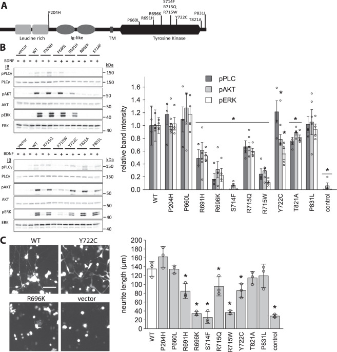
Figure 2.
Functional characterisation of TrkB mutants. (A). Schematic representation of TrkB protein with rare variants indicated; Immunoglobulin (Ig)-like domain; TM (transmembrane region). (B). PC12 cells were transiently transfected with WT/ mutant TrkB. Phosphorylation of downstream signalling molecules was measured by Western Blot before (−) and after (+) stimulation with recombinant BDNF; band intensities quantified by densitometry (right panel; data point = signal from one replicate). Mutants are expressed relative to WT. *p < 0.05, student’s t-test. (C). PC12 cells were transiently transfected with WT/mutant TrkB as well as GFP and stimulated with recombinant BDNF for 48 h. Neurite length was measured by fluorescent microscopy (representative images from 2 mutants shown in left panel; remainder shown in Figure S2) and quantified as length per nucleus (right panel; data point = mean of one replicate). Scale bar: 50 μm. *p < 0.05, student’s t-test.

