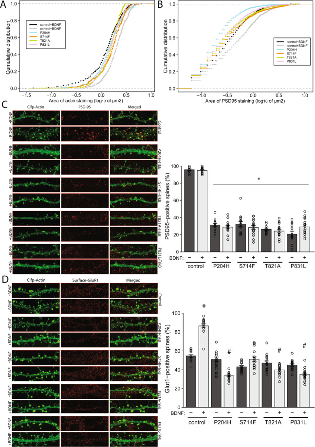
Figure 4.
Effects of TrkB mutants of on dendritic spine maturation. (A). To determine the effects of TrkB mutants on dendritic spines size, rat hippocampal neurons - transiently transfected with Clover fluorescent protein-actin and WT/mutant TrkB - were analysed for spine area before (−) and after (+) recombinant BDNF on DIV11. Representative images are shown in Figs. 3C and 4C (images used for quantification). Cumulative frequency plots for control + /− BDNF and mutants + BDNF are shown. In control, stimulation with BDNF (grey vs black), significantly increases the size of spines. p < 2.2*10−16, Kolmogorov–Smirnov (K-S) test. Expression of mutant forms of Trkb stunts BDNF’s ability to promote spine growth (colours vs grey). p < 0.001, K-S test. (B). To determine the effect of TrkB mutants on post synaptic density sizes, rat hippocampal neurons were transiently transfected with ClFP-Actin and WT/mutant TrkBs (DIV5) and stimulated with vehicle or BDNF (DIV7-11), followed by staining for PSD-95. Representative images are shown (Fig. 4C). Cumulative frequency plots for control + /− BDNF and mutants + BDNF are shown. In control, stimulation with BDNF (grey vs black), significantly increases the area if PSD-95 staining. p < 3*10−15, K-S test. Expression of mutant Trkb diminishes BDNF’s stimulatory effect on postsynaptic density size (colours vs grey). p < 2.2*10−16, K-S test. (C). PSD-95 staining of dendritic spines (mushroom and stubby) is shown in the left-hand panel and proportion of spines positive for PSD-95 staining following expression of WT/mutants is shown in the right-hand panel. PSD-95 staining is reduced for all mutants in the stimulated and unstimulated state. *p < 0.01, student’s t-test ± SEM. (D). To determine the effects of TrkB mutants on the surface expression of GluR1-containing AMPA receptors in dendritic spines, rat hippocampal neurons were transiently transfected with ClFP-actin and WT/mutant TrkBs, followed by live staining for GluR1 in the presence or absence of recombinant BDNF (DIV7-11). Representative images are shown (left-hand panel) and the percentage of dendritic spines (mushroom and stubby) positive for surface GluR1 staining is shown in the right-hand panel. In control, stimulation with BDNF significantly (*p < 0.01, student’s t-test ± SEM) increases GluR1 staining in spines, but shows the reverse effect (i.e. decrease) in 3 of the 4 mutants tested (#p < 0.05, student’s t-test ± SEM).

