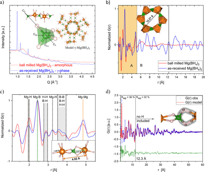
Figure 1.
(a) SR-PXD of γ-Mg(BH4)2 (blue curve) and amorphous-Mg(BH4)2 (red curve). λ = 0.2072 Å. Inset in the upper left is showing three [BH4] tetrahedra in their respective Mg setting and a magnification into one tetrahedron and its rotational axes are shown. C3 is the 3-fold 120° axis and C2∥ and C2⊥ are the 2-fold 180° axis. The inset image shows the crystal structure of γ-Mg(BH4)2 with one interpenetrating channel as reported in ref. 19. Spheres in orange: Mg-, green: B- and grey: H-atoms. (b) PDF obtained from total scattering data collected at P02.1 (DESY) of amorphous and crystalline γ-Mg(BH4)2. λ = 0.20723 Å, inset: One 1D channel of the structure with 12.27 Å diameter. (c) Peaks of the local structure of the amorphous PDF agree well with the crystalline one up to ~5.1 Å. The last coinciding peak is the Mg – Mg distance which is marked in orange in the figure and the structural model inset. The most intense peak corresponds to the Mg-B bond (green). (d) Real space Rietveld fitting of the PDF of γ-Mg(BH4)2. The PDF was fitting using two different models. Details can be found in the text. Inset: Sketch of a three-centre-two-electron-bond.

