
FIGURE 2.
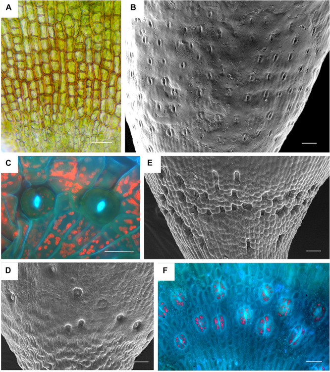
Stomata diversity in mosses. (A) Atrichum angustatum light micrograph of stomata free epidermis. (B) Funaria hygrometrica SEM of apophysis covered with ∼200 stomata. (C) Physcomitrium (Physcomitrella) patens 2 of 10 stomata in fluorescence. (D) Brachythecium rutabulum SEM of sparse scattered stomata. Image credit: Jeffrey J. Duckett. (E) Plagiomnium cuspidatum SEM showing numerous sunken stomata on the apophysis. 60 stomata estimated in the capsule. Image credit: Jeffrey J. Duckett. (F) Bartramia pomiforme group of stomata in fluorescence. 70 stomata estimated in the capsule. Bars: (A,C,F) = 20 μm, (B,D,E) = 50 μm.