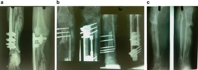
Fig. 4.
a Anteroposterior and lateral radiographs of 25-year-old male patient at the time of presentation showing severe comminution of tibial diaphysis with external fixator in situ. b Immediate postoperative anteroposterior and lateral radiographs of same patient showing 9 cm bone gap with distal corticotomy. c Two year follow-up radiographs showing union at fracture site and consolidation of regenerate. Patient had 3 cm LLD with infection and had fair bony outcome and good functional outcome according to ASAMI criteria

