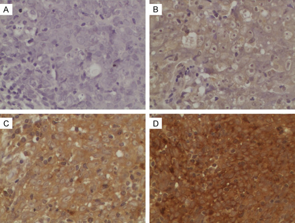
Figure 2.

Representative IHC staining of INHBA in NPC tissue samples. INHBA expression was mainly localized in the cytoplasm. Magnification is 400×. A. Negative staining of INHBA. B. “+” (weakly positive) expression of INHBA. C. “++” (positive) expression of INHBA. D. “+++” (strongly positive) expression of INHBA.
