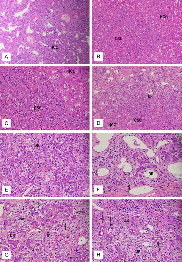
Figure 2.

Histopathologic features of the liver tumor. (A) Hepatocellular carcinoma (HCC) area showing typical compact and trabecular patterns. Tumor vessels are present. H&E, ×40. (B, C) The area of cluster of small cells (CSC) shows an aggregate of small oval cell-like cells with scant cytoplasm, increased nucleo-cytoplasmic ratio, nucleoli, and neutrophilic infiltrations. (B) ×40. (C) ×100. (D, E) The areas of fibrous septa of HCC showing malignant ductular reaction (DR), clusters of small cells (CSC), and HCC cells. There are gradual transitions between HCC cells and CSC cells and also between CSC cells and ductular cells. (D, E) ×100. (F, G) The areas of ductular hepatocytes (arrows) in fibrous septa of HCC showing a mixture of ductular cells and ductular hepatocytes (arrows) that are present singly (F) or in clusters (G). There is a gradual transition between the ductular cells and ductular hepatocyte (F, arrow). (F, G) ×100. (H) The area of fibrous septa of the HCC showing bile duct differentiation (arrows) and ductules (DR). (H) ×100.
