Figure 1.
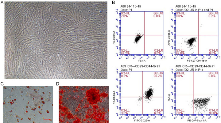
ME-MSC culture and identification. A. ME-MSCs displayed spindle-shaped and fibroblast-like morphology under the microscope. (magnifcation, ×100). B. Identification of ME-MSCs cell surface markers by flow cytometry. ME-MSCs were negative for CD34, CD11b, and CD45, but positive for CD29, CD44, and Sca-1. C. ME-MSCs could respectively in vitro differentiate into osteocyte detected through alizarin red S staining. D. In vitro differentiate into adipocyte detected by oil red O staining.

