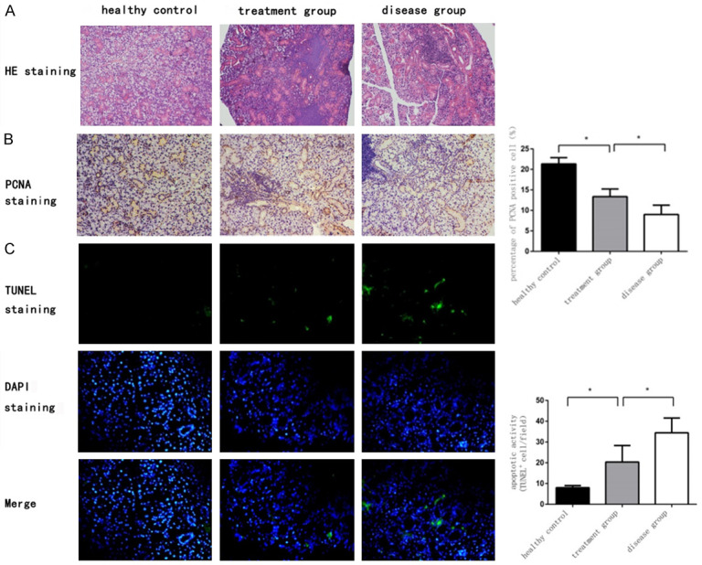
Figure 3.

ME-MSC treatment could restore the acini and micromorphologies, promote the SGEC proliferation, and suppress the SGEC apoptosis from NOD mice. A. HE staining of submandibular glands in mice (magnification, ×200). Relatively well preserved acini and micromorphologies could be found not in the disease group, but in the health control group and in the treatment group. B. PCNA immunohistochemical staining in SGEC in health control group, treatment group, and disease group. The percentage of proliferation cells in disease group [(9.0±2.3%), n=9] was lower than that of treatment group [(13.4±1.9%), n=11]. *P<0.05. C. Apoptosis cells in salivary glands were stained by TUNEL fluorescence staining in health control group, treatment group, and disease group. The percentage of apoptosis cells in treatment group [(20.4±7.9/field), n=11] was lower than that in disease group [(34.4±7.1/field), n=9]. *P<0.05.
