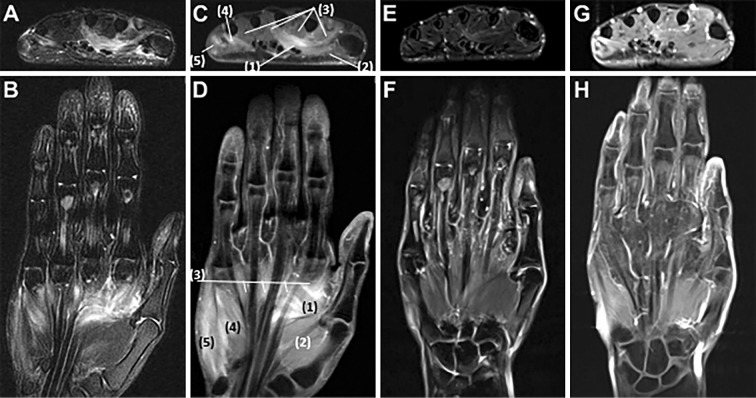
Figure 2.
Magnetic resonance imaging of the left hand. Axial and coronary fat suppression T2-weighted images show edematous changes of the intrinsic muscles of the hand such as the abductor pollicis, abductor digiti minimi, and interosseous muscles (A, B). Axial and coronal contrast T1-weighted images show contrast enhancement of these muscles (C, D). Fat-suppressed T2-weighted images (E, F) and contrast T1-weighted images (G, H) taken 13 days after IVIg treatment show improvement in these inflammatory changes. (1) adductor pollicis, (2) abductor pollicis brevis, (3) lumbricals, (4) flexor digiti minimi brevis, (5) abductor digiti minimi.

