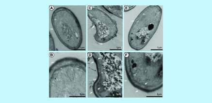
Figure 6. . Transmission electron microscopy of Scedosporium boydii cells incubated for 12 h in the presence of myriocin (16 μg/ml) or PPMP (128 μg/ml).
Control cells were grown in RPMI 1640 supplemented with 2% methanol. After the incubation time, the samples were processed as described in the methodology section and visualized using a ZEISS 900 microscope. White arrows indicate altered membrane (m) and cell wall (cw). (A & B) Control. (C & D) Myriocin. (E & F) PPMP.
PPMP: DL-threo-1-Phenyl-2-palmitoylamino-3-morpholino-1-propanol.

