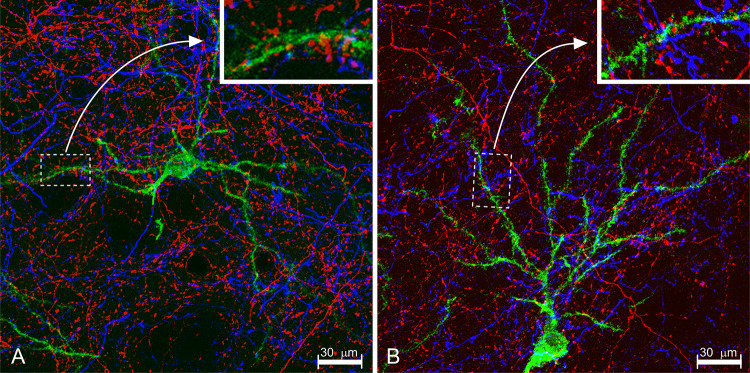
Fig. 7.
Multi-dimensional experiment combining in a rat dual-anterograde tracing with PHA-L (blue) and BDA (red) with retrograde tracing using rabies virus (RV; green). Here, we wanted to evaluate whether corticostriatal and thalamostriatal projections converge onto striatofugal neurons. PHA-L and BDA were iontophoretically delivered into the primary motor cortex and parafascicular thalamic nucleus, respectively. RV was pressure-delivered into either the external globus pallidus or in the substantia nigra pars compacta. a Striatopallidal; B striatonigral neuron. The insets show details. Detection of RV was carried out with an antibody against a soluble rabies phosphoprotein, resulting in Golgi-silver impregnation like labeling of dendrites, with details like dendritic spines

