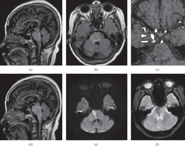
Figure 1.

Magnetic resonance imaging. (a, b) Fluid-attenuated inversion recovery (FLAIR) imaging showing widespread hyperintensity diffusely involving the right pons on day 1. (c) Magnetic resonance angiography showing an abnormal flow signal (arrow) posterolateral to the cavernous sinus. Arrowhead indicates the right internal carotid artery. (d) FLAIR imaging. Brainstem edema has worsened and expanded rostrocaudally on day 2. (e) Diffusion-weighted showing shows hyperintense lesion within the edematous pons. (f) T2 star-weighted imaging showing an area of low intensity, suggesting venous congestion and hemorrhage.
