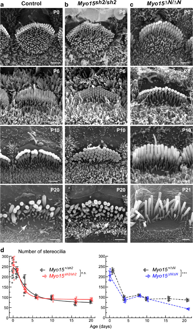
Fig. 2.
Myosin-XVa is not essential for developmental retraction of supernumerary stereocilia in IHCs. a–c SEM images of control heterozygous (Myo15+/sh2) (a), mutant Myo15sh2/sh2 (b), and mutant Myo15∆N/∆N (c) IHC bundles at different postnatal ages, P0-P21. Scale bars: 1 μm. Arrows point to the examples of stereocilia-like projections remaining at the cuticular plate surface of control and Myo15sh2/sh2 IHCs at P20. d Total number of first, second, and third row stereocilia plus stereocilia-like projections in the IHC bundles of Myo15sh2/sh2 (red, left), Myo15∆N/∆N (blue, right), and control heterozygous mice of the same strains (black). Each data point represents individual IHC and is artificially plotted with a small offset relative to the vertical whiskers with a central mark that shows mean ± standard deviations (SDs). All cells were located in the middle of the cochlea. Each time point combines the data from two to three independent biological samples (mice) of each genotype. Asterisks indicate statistical significance of the differences of stereocilia numbers between the rows (two-way ANOVA: n.s., non-significant; *P < 0.05; **P < 0.01; ***P < 0.001). Data for Myo15sh2 strain were fitted to the logistic function, y = A2 + (A1-A2)/(1 + (x/x0)^p), with the following parameters (±SD): Myo15+/sh2 IHCs, A1 = 245 ± 43, A2 = 67.8 ± 14.6, x0 = 2.87 ± 1.33 days, p = 1.65 ± 0.98; Myo15sh2/sh2 IHCs, A1 = 254 ± 13, A2 = 89.1 ± 2.6, x0 = 2.12 ± 0.26 days, p = 2.52 ± 0.43. Unfortunately, we did not have enough data for a reliable fit to the logistic function for Myo15ΔN strain

