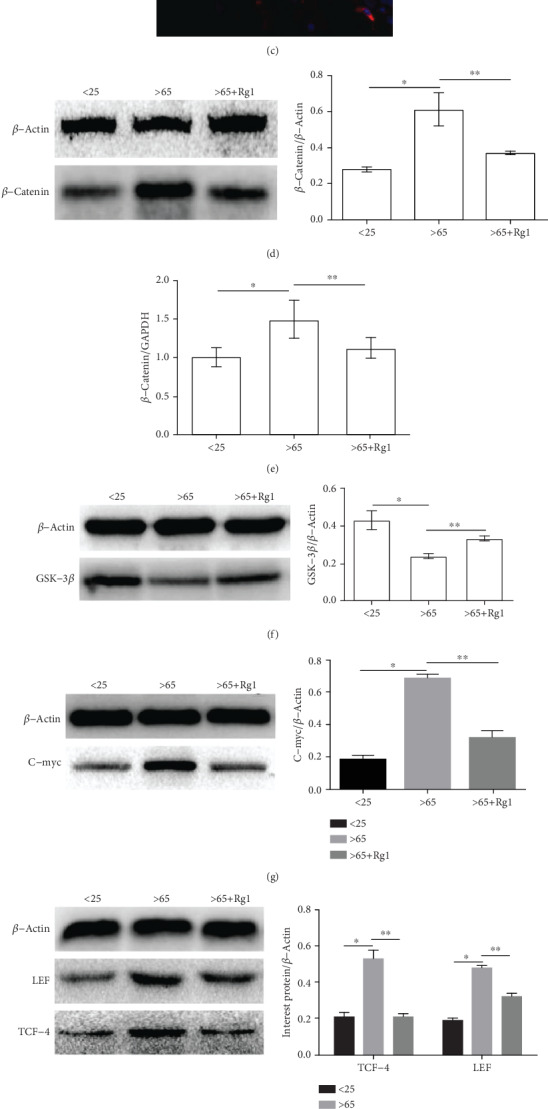
Figure 6.

Effect of Rg1 on the expression of Wnt/β-catenin signaling pathway-related proteins and mRNA in hBM-MSCs. (a–c) By immunofluorescence, PI (red) cells localize β-catenin, DAPI (blue) to visualize the nucleus. (a) the <25-year-old group, (b) the >65-year-old group, and (c) the >65-year-old+Rg1 group. This test was repeated three times. Representative images were shown. (d) Analysis of protein expression of β-catenin by Western blot. β-Actin was used as an internal control. (e, i) Analysis of mRNA of signaling pathway-related mRNA by RT-PCR; mRNA expression of GAPDH was used as an internal control. (f) Expression of GSK-3β protein was analyzed by Western blot. (g, h) Relative protein expression of LEF/TCF and C-myc proteins in different groups, and β-Actin was used as an internal control. ∗∗P < .05 vs. the >65-year-old group, ∗P < .05 vs. the <25-year-old group.
