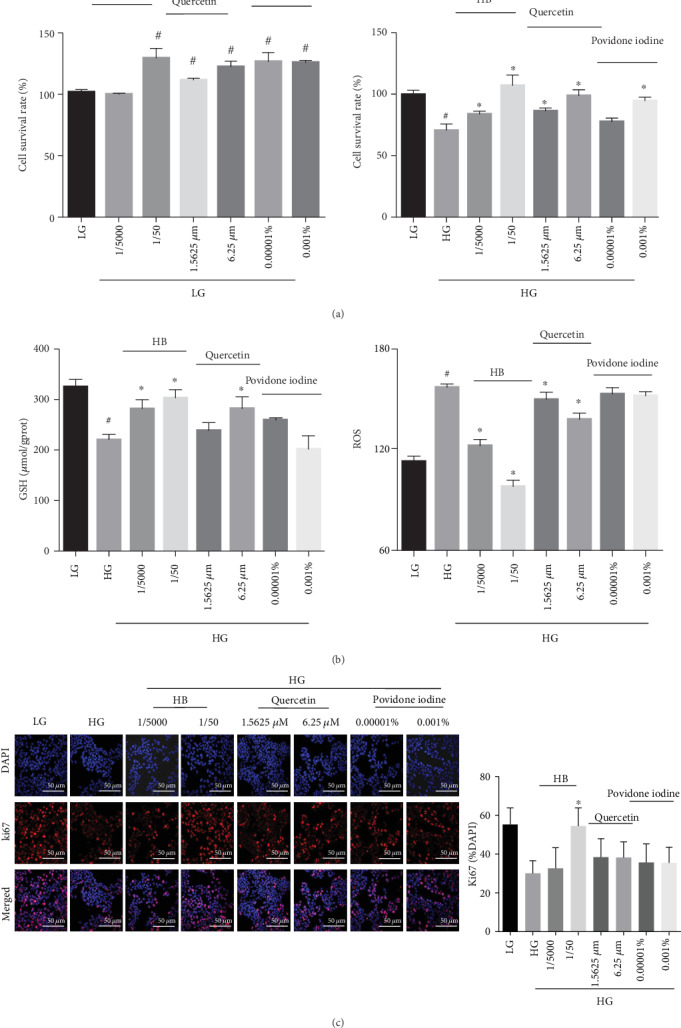
Figure 9.

HB facilitated cell proliferation and reduced oxidative damage in HaCaT cells. (a) Cell viability in LG and HG (n = 6); (b) the levels of GSH (n = 3) and ROS (n = 6); (c) immunofluorescence staining of Ki67 (red) and its quantitative result (n = 3 − 5 per group), nucleus (blue), scale bar: 50 μm; the data were expressed as mean ± SD, and significance was expressed as #P < 0.05 vs LG and ∗P < 0.05 vs HG.
